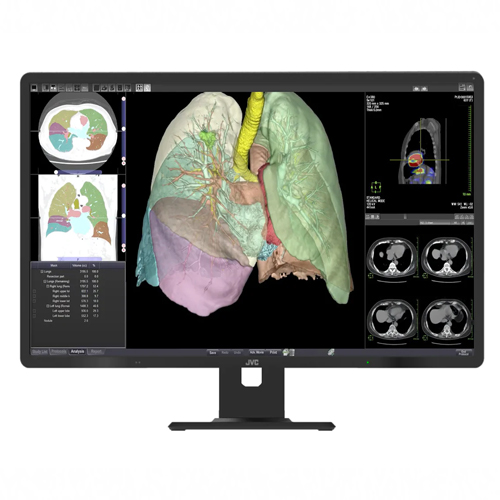
- 21.3-inch, 5 MP Super High-bright LCD Color Diagnostic Display
- 5 Megapixel - 1150 cd/m² - 1800 :1 contrast
- DICOM calibration
- Remote calibration and management
- Color and brightness uniformity
-
-
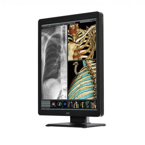
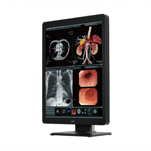
High-bright 3MP color display for diagnostic imaging
Totoku now known as JVC CLS-300 (CLS300) is an optimal solution where doctors need to view both grayscale and color images, and effectively covers multiple applications like PACS, CAD, PET and nuclear medicine.
-
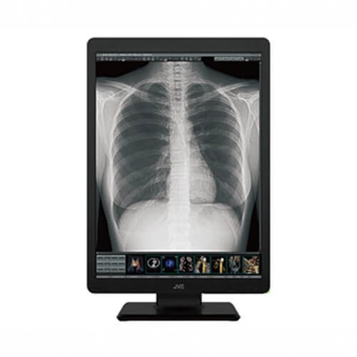
High-bright 2MP color display for diagnostic imaging
Totoku now known as JVC CLS-200 (CLS200) is an optimal solution where doctors need to view both grayscale and color images, and effectively covers multiple applications like PACS, CAD, PET and nuclear medicine.
-
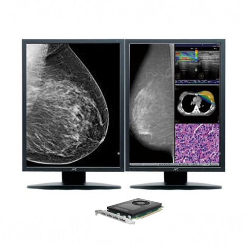
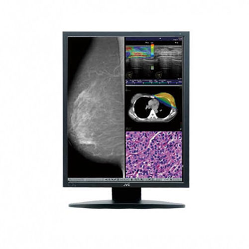
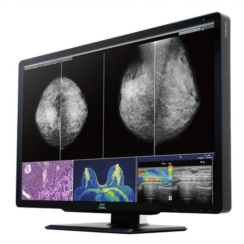 JVC CL-S1200 (JVC CLS1200) can accommodate images from any modality including Digital Mammography/Tomosynthesis, US, CT, MRI and pathology.
- Visual Point Mode
- 1200 cd/m2
- Turbo Luminance
- Dynamic Range Extension
- Protective Glass Filter
- Reading Light & Rear Light
- LED Indicator/Front Buttons
- Biuld-in Sensor
- DICOM Conformance Check
-
- 8MP (3840 x 2160) for multi-modality diagnosis
- Auto Calibration (built-in front sensor)
- Uniformity Equalizer
- Integrated QA Solution
- Built-in Sensor
- Space Saving
- Wire Management / DisplayPort Daisy Chains
-
- Dynamic Gamma
- Integrated QA Solution
- Self-calibration
- LED Indicator / Front Buttons
- Built-in Sensor
-
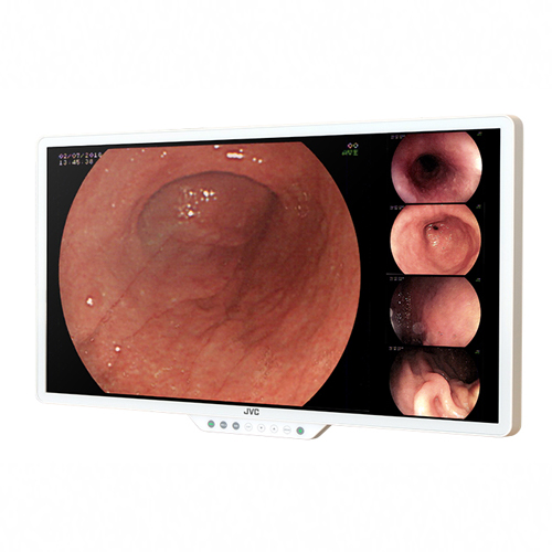
- Full HD 27" Color Monitor
- Widescreen
- Flat design
- 300 cd/m2
- AR Front Glass
- Calibration Function
- Multi-Source
-
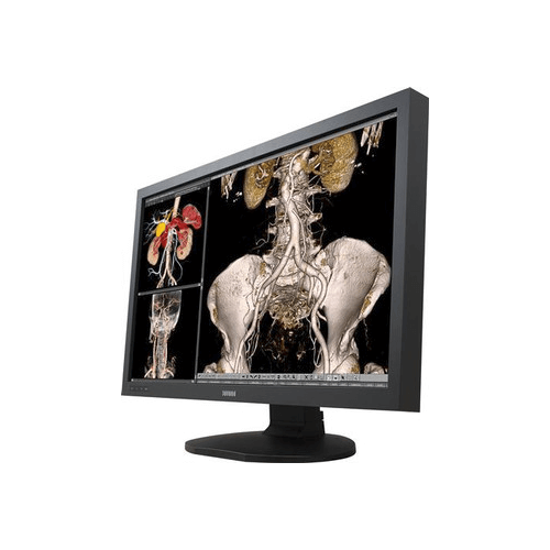
Wide screen 6MP color diagnostic display for new possibilities with streamlined workflow
With a wide 30" screen, the Totoku CCL650i2 allows you to view images of two 3MP displays in one screen without the center bezels. Additionally, the JVC CCL650i2 allows for the flexibility of window layouts in a number of ways supporting multiple system configurations. Providing optimal functions for diagnostic viewing, this seamless and wide LCD improves your workflow efficiency while ensuring space and energy saving.
-
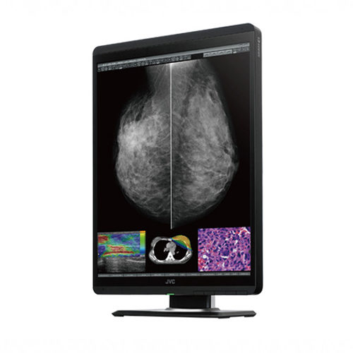
- 21.3-inch, 5 MP High-bright LCD Color Medical Display
- High Resolution and Accommodation Images Monitor for Mammography
- One Pixel Based Contrast and Brightness, Gamma
- Auto Text Mode --- Auto Brightness Blinding Mode for Text Area
- LED Backlight for Image Stability and Energy Saving
- Built-in Sensor
- Remote Calibration
- DICOM Conformance Check
- DVI & DisplayPort
- Uniformity Equalizer
- User-selectable Monitor Configurations
-
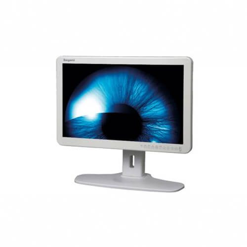
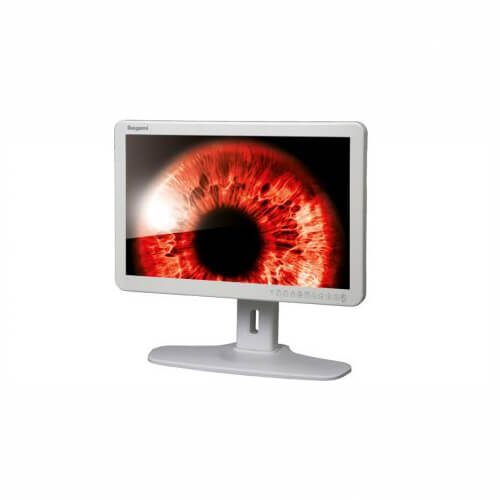
- Fully compliant with medical safety directives
- New Full Flat design for Front panel
- New IPS panel with LED backlight (450 cd/㎡)
- High Resolution, True color display (1.07 Billion Color gradient)
- Jaggy-less I / P Conversion Process
- Multiple Input and Format Capabilities
-
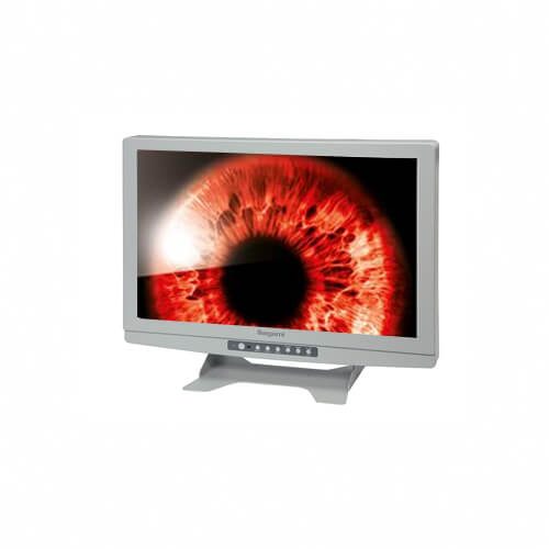
- Fully compliant with medical safety directives
- Wider color gamut than EBU / AMPTE standard (102% NTSC)
- New Full Flat design for Front panel
- New IPS panel with LED backlight (400 cd/㎡)
- High Resolution, True color display (1.07 Billion Color gradient)
- Jaggy-less I / P Conversion Process
- Multiple Input and Format Capabilities
-
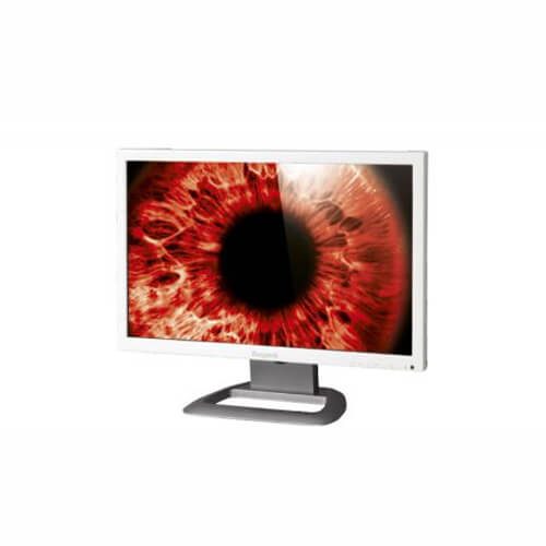
- Fully compliant with medical safety directives
- 21.5-inch Full-HD 3G Multi-Format LED Monitor
- High Resolution Display
- Eco-Friendly LED Solution
- 3D Comb-Filter & De-Interlace
- Slim and lightweight Design
- Multi-adjustable Desktop Stand
-
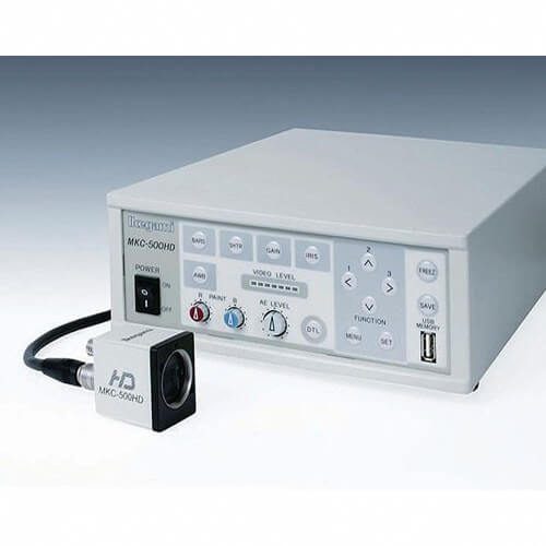 Ikegami MKC500HD (MKC-500HD) 3CMOS FULL HDTV Medical Grade Camera Small size of camera head The camera head employs the compact design on the sensor block with the special optics and achieves the small size and right weight. High quality image The camera equips the latest 1/3-inch CMOS sensors and it performs 1000 TV lines horizontal resolution and 54dB of S /N ratio. High sensitivity The cameras offers the highest sensitivity on HDTV medical grade cameras with F12/2000lx. And it equips the high sensitivity mode and it performs the double of standard sensitivity.
 -
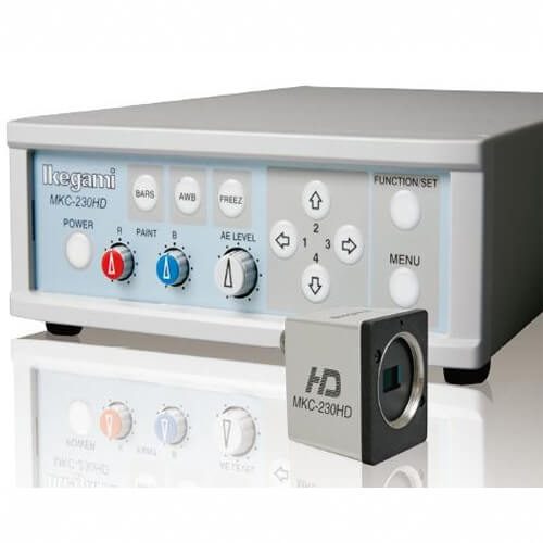
- Resolution 1920 x 1080P
- 3 chip camera
- CMOS sensor
- Outputs: DVI and HD-SDI
- C-Mount camera
-
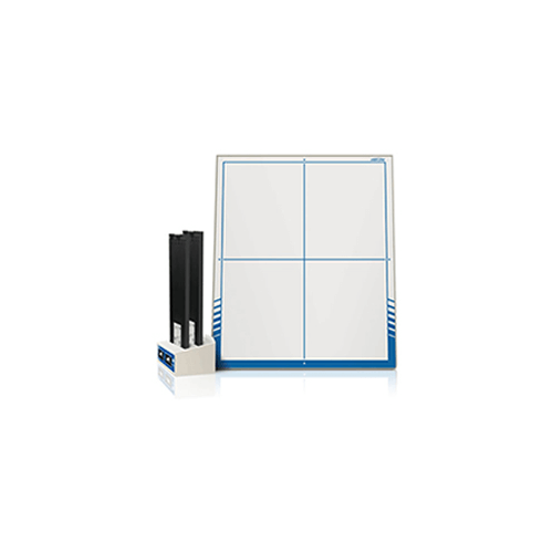
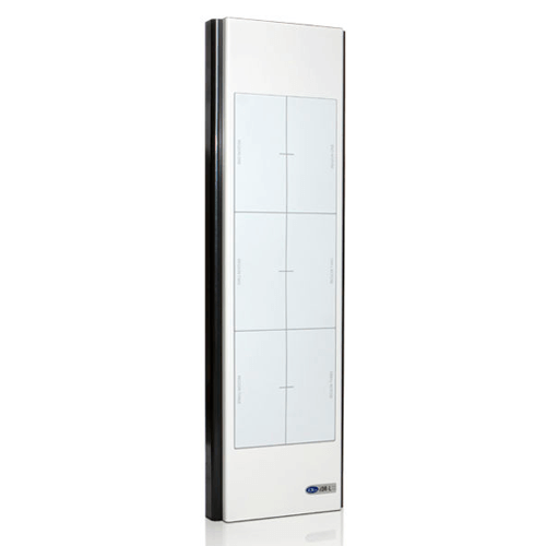 One solution to address all your long-bone imaging needs. Introducing the iDR-L. A seamless, high-resolution detector that offers full spine, regional, and zone scanning capabilities. This direct capture solution revolutionizes how full-spine imaging is achieved. iDR-L Method: The long-bone radiography platform utilizes a 17” by 51” detector plate that eliminates the need for a cassette. Integrating phosphor plate technology, the iDR-L boasts a higher resolution than standard DR while maintaining the same workflow. This solution sends complete image information directly to the scan processor skipping lengthy image stitching processes.
 -
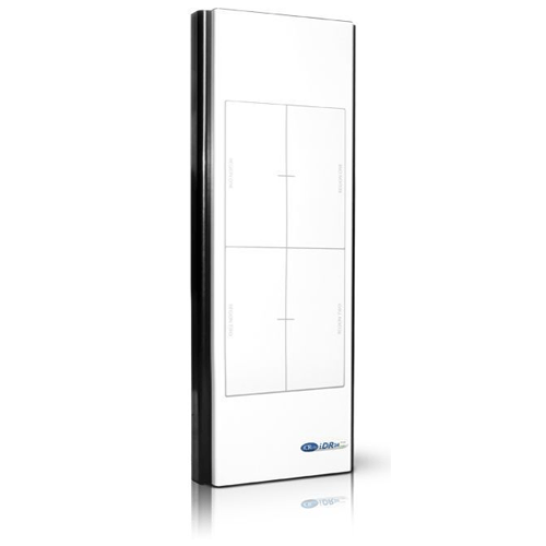 iCRco iDR-34, the only digital radiography solution capable of BOTH regional scanning and long length spinal imaging studies. With a 17”x34” field of use, the iCRco iDR-34 can replace or supplement your existing RAD room environment by giving you the ability to do all anatomy regions - neck and chest, full-torso, long-length, and spine. Because the system takes a single image, you can leverage compensation filters to balance out the exposure between thicker and thinner areas of anatomy.
 -
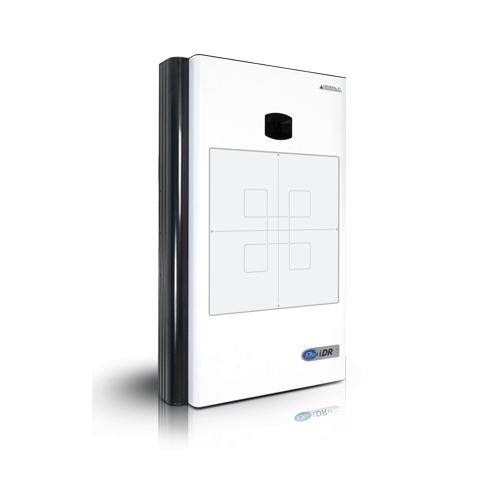 The iCRco iDR boasts a 14x17 scanning area with 17x17 option. This affordable fixed panel deploys a unique scanning DR technology that allows for a direct digital workflow at a much lower cost than standard fixed DR panels. iDR produces amazing image quality and is backed by powerful optics, sensors, and a solid-state, mechanical drive system with only one moving part. Add the optional wall stand for imaging flexibility for chest, abdomen and simultaneous poster anterior (PA) and lateral imaging.
 -
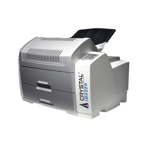
Crystal Clear Images
-
Excellent Reliability, Minimum Maintenance
-
Direct Digital Imaging Technology
-
Convenient Imaging With Two Media Sizes On-Line
-
-
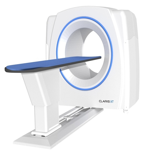
- Blazing Fast Performance
- 20 Second Acquisition
- 2-5 Minutes Reconstruction
- 17" x 17" Sensor Size
- 16 Bit Large Area Dynamic Sensor
- 140 μm Voxel Pitch
-
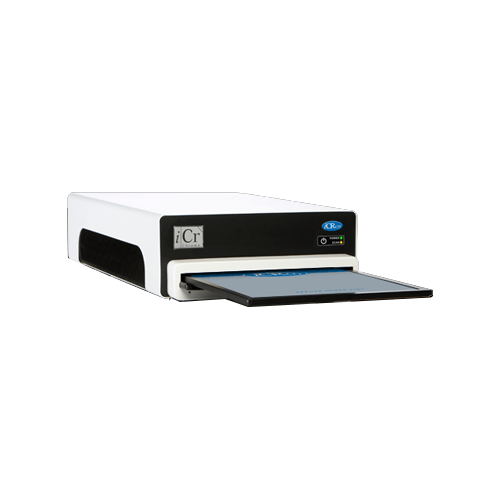
 The Chrome Neo is paired with XC™, iCRco’s superior image acquisition software. Custom settings can be defined based on user preference, allowing you to apply advanced image processing algorithms to look for even the smallest details. Add our complete computed radiography workflow solution and you have the whole package - image acquisition, practice management, PACS, a desktop viewer, a mobile viewer for the iPad™ or iPhone™, an incomparable image wizard, and a comprehensive QC tool. It’s neonatal/ pediatric imaging as you’ve never seen it before.
 -
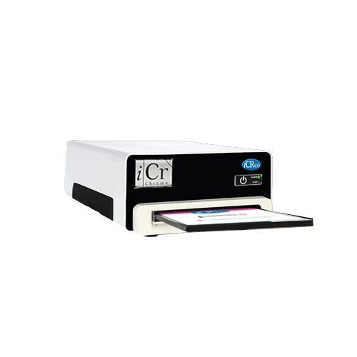
 Small is the new big. Introducing Chrome™, the elemental CR - a lightweight, compact, tabletop scanner designed specifically for the modern practice. This affordable, single-plate reader produces amazing image quality and is backed by powerful optics, sensors, and a solid-state, mechanical drive system. Physicians that require a reliable imaging system can now achieve amazing workflow at a much lower cost than comparable computer radiography solutions..
 -
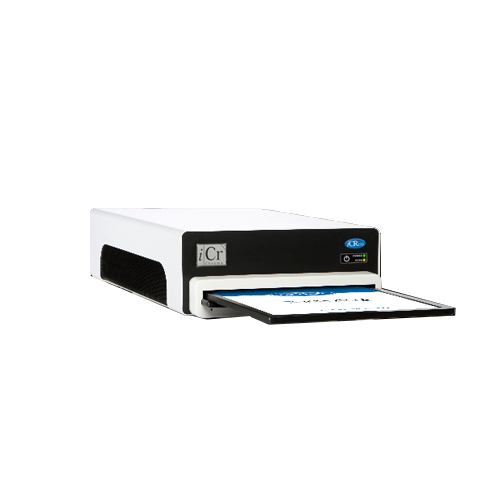 Chrome+ is paired with XC, iCRco’s superior image acquisition software. Custom settings can be defined based on user preference, allowing you to apply advanced image processing algorithms to look for even the smallest details. Add our complete imaging workflow solution and you have the whole package: Image Acquisition, RIS-PACS, a Desktop Viewer, a Mobile Viewer for the iPad.
 -
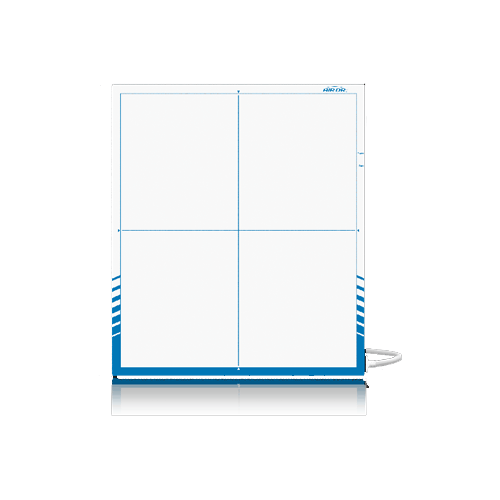 The AirDRc o ffers amazing versatility and lightning-fast image display speed. This cassette-sized, flat panel integrates seamlessly into any new or existing radiology room. It can be utilized for all your table work, and weight-bearing views. With a small upfront investment you can transform your radiology department into an e fficient, profit center with increased patient throughput and a smoother acquisition experience. Get connected to the future with AirDRc.
 -
 The iCRco AirDRc has been specially designed and optimized to advance the imaging equipment you’re using right now. Utilizing it's unique form-factor and embedded Automatic Exposure Detection (AED), the AirDRc is compatible with any X-ray system that works with ISO 4090 - compliant, 43 x 43 cm cassettes.
- No need to modify your generator, or bucky
- No need to replace your grids
- No need to discard your wall stand, or table.
- Water and Dust Resistant IP-65 Rating
- Carbon Fiber Construction Durability
- Same Day Installation
-
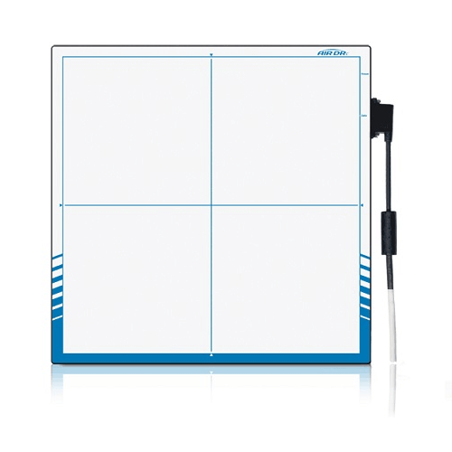
 The iCRco AirDR System has been specially designed and optimized to advance the imaging equipment you’re using right now. Utilizing it's unique form-factor and embedded Automatic Exposure Detection (AED), the AirDR System is compatible with any X-ray system designed to work with ISO 4090 - compliant, 35 x 43 cm cassettes. Innovative Direct Radiography Panel Amorphous Silicon active TFT/diode array, Carbon-fiber construction. Scintillator Direct Deposit: Cesium Iodide Pixel Matrix 3556 × 4320 Pixel Pitch 100 μm Image Data 16 bit Image Transfer Time Wired: 500 ms; Wireless: 3000 ms Active Area True 35.5 cm × 43.2 cm External Dimensions ISO 4090 cassette size 14” × 17" (38.4 cm (w) × 46 cm (l) × 1.5 cm (h)) Weight 8.4 lbs (3.8 kg) Status Display LED display (Wifi/Battery/Sensor) Wireless Data I/F 802.11n WiFi standard Wired Data I/F GigE via optional power & communication tether X-ray I/F Automatic Exposure Detection (AED) Limiting Resolution 5 lp/mm Typical MTF 70% (1 lp/mm), 40% (2 lp/mm), 15% (4 lp/mm) for RQA5 Typical DQE 75% (0 lp/mm), 60% (1 lp/mm), 40% (3 lp/mm) for RQA5 Environment 10 – 35 °C operating, 30 – 70 % RH operating (non-condensing) Battery Rechargeable battery, 53.3 Wh Battery Charger External two bay charger 100-240 V AC 50/60 Hz Interface and Power Unit Optional AirDR IPU with external power supply 100-240 V AC, GigE, and X-ray I/F Standards IEC 60601-1, IEC 60601-2, IEC 60601-1-6, FCC 47CFR PT 15, FCC OET 65C, ETSI EN 301 893, EN 62311, ISO 10993-5, ISO 10993-10, CE Binned Mode (option) Up to 8 fps for 2 x 2 binned, 200 μm pitch for a pixel matrix of 1778 x 2160" Image Calibration (option) On-board offset, gain and defective pixel corrections Fast Preview (option) 4 x 4 binned quick preview image WAP Mode (option) Wireless Access Point functionality.
- No need to modify your generator, or bucky
- No need to replace your grids
- No need to discard your wall stand, or table.
- Water and Dust Resistant IP-65 Rating
- Carbon Fiber Construction Durability
- Same Day Installation
-
- End of Life Model
- Limited availability
- Available on Exchange
- Repair Service Available
-
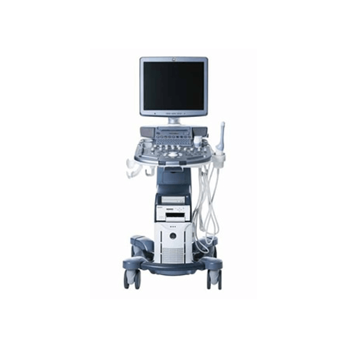 The Voluson S8 system opens new opportunities in clinical imaging while enabling the efficiency and productivity that busy practices demand. Among its advantages:
- Extraordinary image quality — the foundation of Voluson ultrasound— to help achieve a clear view into obstetric and gynecologic exams.
- Sophisticated fetal assessment tools — to help support earlier, more detailed evaluations.
- Innovative probe technologies — to support thorough evaluations of even the most challenging patients.
- Easy imaging — with system intelligence and probe technology combining to produce outstanding images with minimal user interaction.
- Easy-to-use automation tools — that help streamline workflow, forge stronger connections with patients and referring physicians, and control costs.
- Ergonomic design — that simplifies how users interact with the system and helps provide optimal comfort while scanning.
-
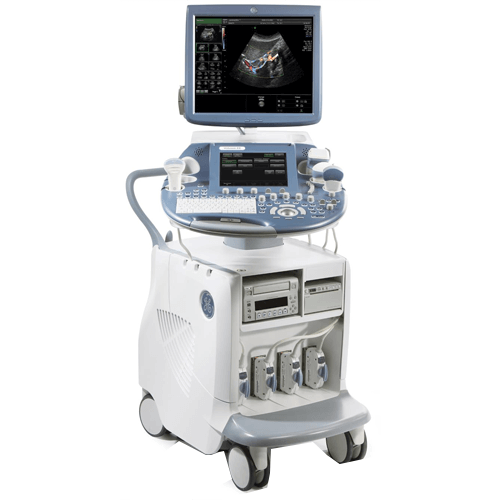
- Touch screen — the user interface helps enhance workflow speed by eliminating keystrokes and displaying critical functions prominently.
- New PDF export capabilities — This tool allows the export if report data in PDF format with just the push of a button.
- Virtual Rescan — The system captures raw data with every exam, enabling a virtual rescan of the patient at any time after the study, on the system or at a remote ViewPoint workstation.
- Anatomical M-Mode (AMM) — Apply M-Mode to two separate areas of the fetal heart to help assess arrhythmias—either while scanning real-time or from a stored clip.
- Scan Assistant — Customizable and user-friendly tool that helps improve quality assurance, increase exam consistency, and enhance productivity.
- DICOM1 — compliant and compatible with many electronic medical record systems
- ViewPoint — reporting and image management enables clinicians to create electronic reports that include images and charts, enhancing archiving and physician communication. ViewPoint also enables volume manipulation with 4DView option, and ultrasound data importation to your medical record system.
-
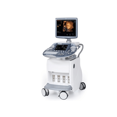 The powerful GE Voluson E6 is designed specifically to give you the exceptional vision you need today so you have the flexibility to meet your emerging needs. This versatile system helps deliver expandable capabilities to grow with your practice. Voluson E6 has enhancements to help you improve patient care and simplify your workflow.
 -
- 4x the processing power of the Voluson e10
- 4D Electronic Matrix 4D Transducer
- HD Live
- V-SRi advanced volumetric speckle reduction
- 4D Electronic Matrix 4D Transducer
- 23” Widescreen LED monitor
- 12” multitouch touch panel
- Probe port illumination
- High frame rates in 2D/4D imaging
- Ergonomically designed for maximum comfort